
Cats may look very different from us on the outside, but underneath all that fur, their basic anatomy is surprisingly familiar. Like humans, cats are mammals with the same major organ systems, including the digestive, respiratory, circulatory, and urinary systems.
That said, cats are not simply small versions of humans. Their organs are adapted to their own diet, lifestyle, and evolutionary history, and some of their physiology works quite differently from ours.
In this guide, we’ll take a closer look at the major internal organs in a cat, where they are located, what they do, and some of the health problems that can affect them.
A Diagram of Cat Internal Anatomy
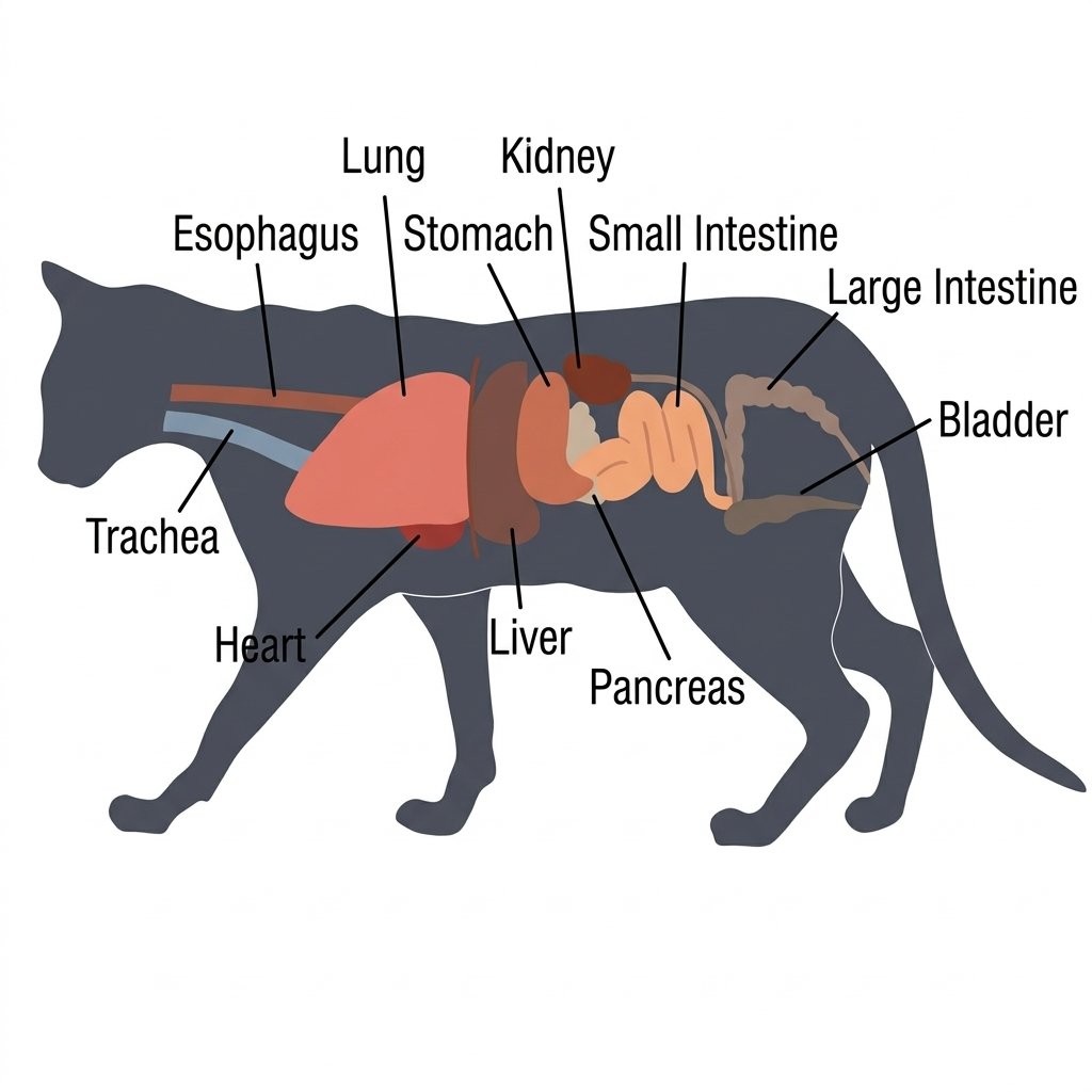
A cat’s internal organs work together as interconnected systems rather than as isolated parts. Many involuntary functions such as heart rate, digestion, breathing patterns, and some aspects of urination, are regulated by the autonomic nervous system.
The autonomic nervous system has two main branches: the sympathetic nervous system and the parasympathetic nervous system.
The sympathetic nervous system becomes more active during exercise, fear, injury, and other stressful situations. This is often described as the body’s fight-or-flight response. The parasympathetic nervous system generally becomes more active during rest and helps the body return to a calmer state.
Together, these systems help regulate many basic functions, including circulation, digestion, breathing, body temperature, and other involuntary processes.
The Cat Digestive System
A cat’s digestive system is responsible for taking in food, breaking it down into usable nutrients, absorbing those nutrients, and eliminating what the body cannot use.
It includes the mouth, esophagus, stomach, liver, pancreas, small intestine, large intestine, rectum, and anus. Digestion begins in the mouth and continues as food moves through the gastrointestinal tract.
A Cat’s Mouth
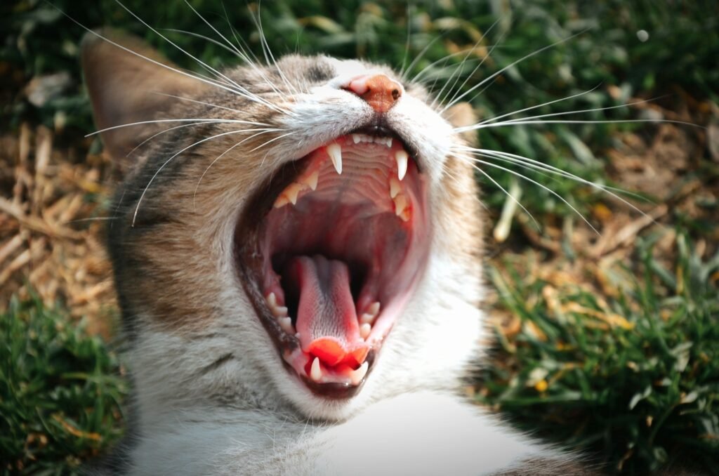
Digestion starts as soon as a cat picks up food with its mouth. The teeth break the food into smaller pieces, while the tongue helps move it around the mouth and prepare it for swallowing.
Once the food is swallowed, it is formed into a soft mass called a bolus, which travels down the esophagus toward the stomach.
One of the more interesting features of a cat’s mouth is the hard palate, or the bony roof of the mouth. Its surface contains raised folds known as palatal rugae.
Cat owners are probably more familiar with another unusual feature: the rough surface of the tongue. A cat’s tongue is covered with tiny, backward-pointing structures called filiform papillae. These keratinized, spine-like structures help cats grip and manipulate food. Studies of feline tongues have documented several distinct types and arrangements of these papillae.
Cats also have palatal rugae, although their number varies by species. One commonly cited comparison is:
| Animal | Typical number of palatal rugae |
|---|---|
| Horse | 16–18 |
| Cow | 15–20 |
| Sheep | 14 |
| Goat | 12 |
| Pig | 20–22 |
| Dog | 9 |
| Rabbit | 15 |
| Cat | 7 |
In cats, individual palatal ridges may also contain small projections that point toward the back of the mouth.
The original description of this structure compares it with structures found in the mouth of leatherback sea turtles. While the similarity is interesting, claims about exactly why these structures evolved should be treated as evolutionary hypotheses rather than established fact.
A Cat’s Esophagus
The esophagus is a muscular tube that carries swallowed food from the throat to the stomach.
As food travels downward, it passes through an opening in the diaphragm called the esophageal hiatus before reaching the stomach.
The esophageal wall contains several layers, including the mucosa, submucosa, and muscular layer. The lining helps protect the esophagus and provides lubrication, while coordinated contractions of the muscular wall push food toward the stomach.
This wave-like movement is called peristalsis.
Problems involving the esophagus can interfere with the normal movement of food. Esophagitis refers to inflammation of the esophagus, while megaesophagus describes an abnormally enlarged esophagus. Esophageal achalasia is a specific disorder involving abnormal relaxation at the lower end of the esophagus.
A Cat’s Stomach
The stomach is a muscular organ that temporarily stores food after it leaves the esophagus. It also mixes food with gastric secretions and begins the digestion of proteins before the partially digested material moves into the duodenum, the first section of the small intestine.
The stomach wall contains several layers, including the mucosa, submucosa, and muscular layer.
The stomach lining contains countless tiny openings called gastric pits. These lead to gastric glands that produce substances including hydrochloric acid and pepsinogen, an inactive precursor of the digestive enzyme pepsin.
Once activated, pepsin helps break down proteins into smaller molecules.
The stomach has several important jobs:
- Temporarily storing food after it is swallowed
- Mixing food with gastric secretions
- Using stomach acid to create an environment that supports digestion and helps limit the growth of many microorganisms
- Beginning significant protein digestion with pepsin
- Producing hormones and signaling molecules, including gastrin, that help regulate gastric activity
When the stomach becomes inflamed, the condition is called gastritis. An ulcer in the stomach lining is called a gastric ulcer. A twisting of the stomach is known as gastric torsion or gastric dilatation-volvulus (GDV), although GDV is much more common in dogs than cats.
A Cat’s Liver
The liver is a large organ located just beneath the diaphragm. It performs a remarkably wide range of jobs and is central to digestion, metabolism, nutrient processing, and detoxification.
Some of its major functions include:
- Processing carbohydrates
- Metabolizing fats
- Processing proteins and amino acids
- Producing and storing glycogen
- Helping maintain blood glucose levels
- Producing bile
- Converting ammonia into urea
- Processing drugs and other substances
- Producing important blood proteins, including albumin
Cats, like other mammals, can synthesize vitamin C in the liver, so they do not normally need dietary vitamin C to prevent deficiency.
The liver produces bile, which is stored and concentrated in the gallbladder and released into the small intestine when needed for digestion.
Liver disease can take many forms. Hepatitis refers to inflammation of the liver. Excessive fat accumulation in the liver is called hepatic lipidosis, commonly known as fatty liver disease. Long-term damage and scarring of the liver can lead to cirrhosis.
A Cat’s Pancreas
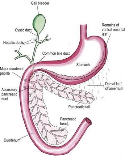
The pancreas plays two very different but equally important roles: digestion and blood sugar regulation.
Most of the pancreas consists of exocrine tissue, which produces digestive enzymes. A smaller portion consists of endocrine tissue, including clusters of cells called the islets of Langerhans.
The exocrine pancreas produces pancreatic juice, which travels through the pancreatic duct into the duodenum. It contains digestive enzymes such as:
- Trypsin and chymotrypsin, which help digest proteins
- Amylase, which helps digest carbohydrates
- Lipase, which helps digest fats
The islets of Langerhans produce hormones such as insulin and glucagon. Insulin helps lower blood glucose by promoting the uptake and storage of glucose, while glucagon helps raise blood glucose when levels are too low.
Inflammation of the pancreas is called pancreatitis. When blood glucose remains abnormally high because the body does not produce enough insulin or cannot use insulin effectively, the result is diabetes mellitus, commonly called diabetes.
A Cat’s Small Intestine
The small intestine connects the stomach to the large intestine and is where most digestion and nutrient absorption takes place.
Its inner lining is specially designed to maximize the surface area available for absorption. The intestinal wall contains folds covered with millions of tiny finger-like projections called villi. Each villus is covered with even smaller structures called microvilli.
Together, these folds, villi, and microvilli dramatically increase the available surface area, allowing nutrients to be absorbed efficiently.
As food moves through the small intestine, carbohydrates, proteins, fats, vitamins, minerals, and other nutrients are broken down into forms the body can absorb. Digestive enzymes attached to the intestinal lining also help complete the final stages of digestion.
Nutrients are then absorbed through the intestinal wall and transported into the bloodstream or lymphatic system.
Material that remains after digestion and absorption continues into the large intestine.
Inflammation of the intestine is called enteritis. A blockage that prevents intestinal contents from moving normally is called an intestinal obstruction.
A Cat’s Large Intestine
The large intestine continues from the small intestine and includes the cecum, colon, and rectum.
In cats, the cecum is relatively small because cats are obligate carnivores and have less need for the extensive fermentation of plant material seen in many herbivores.
The large intestine has several important functions:
- Absorbing water and electrolytes
- Housing large populations of gut bacteria
- Fermenting some undigested material, particularly certain types of dietary fiber
- Forming and storing feces
The rectum connects to the anus, through which feces are eventually expelled from the body.
When feces become difficult to pass or remain in the colon for too long, a cat may develop constipation. An abnormally enlarged colon is called megacolon. When the colon cannot properly absorb water, stools may become loose or watery, resulting in large-bowel diarrhea.
The Cat Respiratory System
The respiratory system brings oxygen into the body and removes carbon dioxide produced by normal metabolism.
Air enters through the nose or mouth and travels through the upper airways before reaching the lungs, where oxygen and carbon dioxide are exchanged.
A Cat’s Trachea
The trachea, commonly called the windpipe, is the airway that carries air from the larynx toward the lungs.
At its lower end, the trachea divides into the two main bronchi, which enter the lungs. Inside the lungs, the bronchi branch repeatedly into smaller airways called bronchioles, which ultimately lead to the alveoli.
At the upper end of the trachea is the larynx, or voice box. The larynx helps protect the lower airway during swallowing and also plays an important role in vocalization.
Cats produce a wide range of sounds using structures within and around the larynx.
Inflammation of the bronchi is called bronchitis. Cats can also develop feline asthma, a chronic inflammatory airway disease that can cause coughing, wheezing, and episodes of breathing difficulty. Asthma should not simply be described as chronic bronchitis; they are related but distinct conditions.
A Cat’s Lungs
Cats have two lungs located inside the chest cavity. The lungs sit above the diaphragm and are surrounded by the rib cage and intercostal muscles.
As the bronchi branch repeatedly inside the lungs, they eventually lead to millions of tiny air sacs called alveoli.
The alveoli are the primary sites of gas exchange. Oxygen from inhaled air passes into the blood, while carbon dioxide moves from the blood into the alveoli and is then exhaled.
A cat’s lungs have a very large total surface area for gas exchange, although estimates can vary depending on how lung surface area is measured.
Breathing depends heavily on changes in pressure inside the chest.
When the diaphragm contracts, it flattens and increases the volume of the chest cavity. The intercostal muscles also help expand the chest. As the chest cavity expands, pressure inside the lungs falls and air flows inward.
During normal exhalation, the respiratory muscles relax and the elastic recoil of the lungs and chest wall helps push air back out.
Diseases affecting the lungs can seriously interfere with this process. Pneumonia is inflammation and infection of lung tissue. Emphysema involves abnormal enlargement and destruction of air spaces, while pneumothorax occurs when air enters the pleural space and causes the lung to partially or completely collapse.
A Cat’s Diaphragm
The diaphragm is a dome-shaped muscle that separates the chest cavity from the abdominal cavity. It plays a central role in breathing.
Several important structures pass through openings in the diaphragm, including the aorta, esophagus, and caudal vena cava. These openings are known as the aortic hiatus, esophageal hiatus, and caval opening.
The diaphragm is controlled by the phrenic nerves, which arise from the cervical spinal cord. This nerve supply is important because the diaphragm can continue to function even when movement of the ribs is impaired by certain spinal injuries, provided the phrenic nerves and diaphragm remain intact.
When the diaphragm contracts, it flattens from its normal dome shape and increases the volume of the chest cavity. This lowers pressure inside the chest and draws air into the lungs.
In simple terms, the diaphragm works much like a bellows, helping pull air into the lungs with each breath.
The diaphragm is one of the most important muscles involved in breathing, working together with the intercostal muscles.
If the diaphragm tears because of trauma and abdominal organs move into the chest cavity, the condition is called a diaphragmatic hernia.
The Cat Circulatory System
The circulatory system moves blood throughout the body, delivering oxygen and nutrients to tissues while carrying carbon dioxide and other waste products away.
The heart is the central pump, while blood vessels provide the network through which blood travels.
A Cat’s Heart
The heart is a muscular pump that keeps blood moving through the lungs and the rest of the body.
Most of the heart is made of cardiac muscle, or myocardium. This muscle contains both working cardiac muscle cells, which contract to pump blood, and specialized cardiac cells that generate and conduct the electrical signals responsible for the heartbeat.
These electrical signals coordinate the heart’s contractions and allow it to beat in a regular rhythm.
A cat weighing about 11 pounds (5 kg) has roughly 11 fluid ounces (330 mL) of blood in its body.
The circulation through the heart and lungs follows a continuous loop:
- Blood with relatively low oxygen levels enters the right atrium.
- It moves into the right ventricle.
- The right ventricle pumps it through the pulmonary artery to the lungs.
- In the lungs, the blood releases carbon dioxide and picks up oxygen.
- Oxygen-rich blood returns to the left atrium through the pulmonary veins.
- It moves into the left ventricle.
- The left ventricle pumps the blood through the aorta to the rest of the body.
- After delivering oxygen to tissues, the blood returns to the heart through the veins and the cycle begins again.
This two-part circulation allows the lungs to oxygenate the blood before the heart sends it throughout the body.
When the heart cannot pump enough blood to meet the body’s needs, the result may be heart failure. Diseases that primarily affect the heart muscle are known as cardiomyopathies.
A Cat’s Blood
Blood is a specialized fluid that travels through the body’s blood vessels. It contains several major components, including red blood cells, white blood cells, platelets, and plasma.
Red blood cells contain hemoglobin, a protein that binds oxygen and carries it throughout the body. In cats, red blood cells are produced mainly in the bone marrow and are removed from circulation as they age, primarily by the spleen and other tissues. A cat’s red blood cells have a shorter lifespan than human red blood cells, averaging around 70–80 days.
White blood cells are part of the immune system. Different types of white blood cells identify and respond to bacteria, viruses, parasites, damaged cells, and other threats.
Platelets help the blood clot. When a blood vessel is damaged, platelets gather at the site and help form a clot that limits blood loss.
Several important diseases can affect the blood. Excessive destruction of red blood cells can cause hemolytic anemia. When the kidneys cannot adequately remove nitrogen-containing waste products, these substances can build up in the bloodstream, resulting in uremia. Leukemia is a type of cancer involving abnormal production and proliferation of blood-forming cells.
The Cat Urinary System
The urinary system removes metabolic waste from the bloodstream and eliminates it from the body as urine.
But that’s not its only job. The kidneys also help regulate water balance, electrolytes, blood pressure, red blood cell production, and other aspects of the body’s internal environment.
The main organs of the urinary system are the kidneys, ureters, bladder, and urethra.
A Cat’s Kidneys
Cats have two kidneys, located toward the back of the abdominal cavity, one on each side of the spine.
The kidneys filter the blood and selectively reabsorb water and useful substances while helping remove metabolic waste into the urine.
The functional units of the kidneys are called nephrons. Each nephron contains structures including the renal corpuscle, which includes Bowman’s capsule and the glomerulus, along with the renal tubules.
The kidneys also perform several important regulatory functions. They release renin, which is involved in blood pressure regulation, and erythropoietin (EPO), which signals the bone marrow to produce red blood cells. The kidneys also play a role in maintaining fluid and electrolyte balance and in processing vitamin D.
Cats are especially efficient at conserving water. Their ancestors, including the African wildcat, evolved in relatively dry environments, and modern cats generally produce concentrated urine.
Urine output varies with factors such as water intake, diet, temperature, activity, and health. For that reason, a single daily urine volume should not be treated as a universal normal value for every cat.
Likewise, protein can sometimes be detected in a cat’s urine without automatically meaning that the kidneys are failing. Persistent or significant protein loss in the urine, however, can be an important sign of kidney or urinary tract disease and should be evaluated by a veterinarian.
Kidney inflammation is called nephritis. When the kidneys lose enough function that they can no longer adequately remove waste products and maintain the body’s internal balance, the result is kidney failure or advanced kidney disease.
Diseases affecting the bladder or urethra and other structures of the lower urinary tract are commonly grouped under feline lower urinary tract disease (FLUTD). FLUTD is a broad clinical term rather than a single disease.
A Cat’s Bladder
The bladder is a muscular, expandable organ that temporarily stores urine.
Urine produced by the kidneys travels through the ureters and enters the bladder. When the bladder fills, its walls stretch. Nerves and the brain coordinate the sensation of bladder fullness and the reflexes involved in urination.
When a cat urinates, the bladder contracts while the urethral outlet relaxes, allowing urine to leave the body through the urethra.
A cat’s bladder does not have one fixed capacity that applies to every individual. Its volume changes depending on the cat’s size, hydration, urine production, and how full the bladder is at a given moment.
Common bladder disorders include cystitis, which means inflammation of the bladder, and bladder stones, which are mineral formations that develop within the urinary tract.
Because urinary problems can sometimes become emergencies in cats—especially when a cat cannot pass urine—changes such as frequent trips to the litter box, straining to urinate, crying while urinating, blood in the urine, or producing little or no urine should not be ignored.