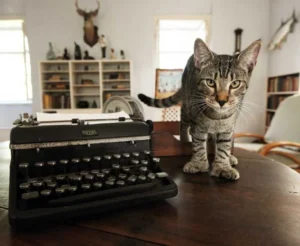
A cat’s skeletal structure is almost identical to that of other big cats like lions, with the main difference being size. In other words, cats have a skeleton that has evolved specifically for a carnivorous lifestyle, perfectly adapted for hunting and agile movement.
In the following sections, we’ll explore the details of a cat’s skeleton.
Cat Skeleton Overview
The domestic cat’s skeletal structure is nearly the same as that of larger cats such as lions and tigers, with the only real difference being scale.
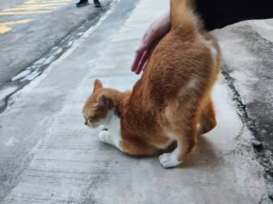
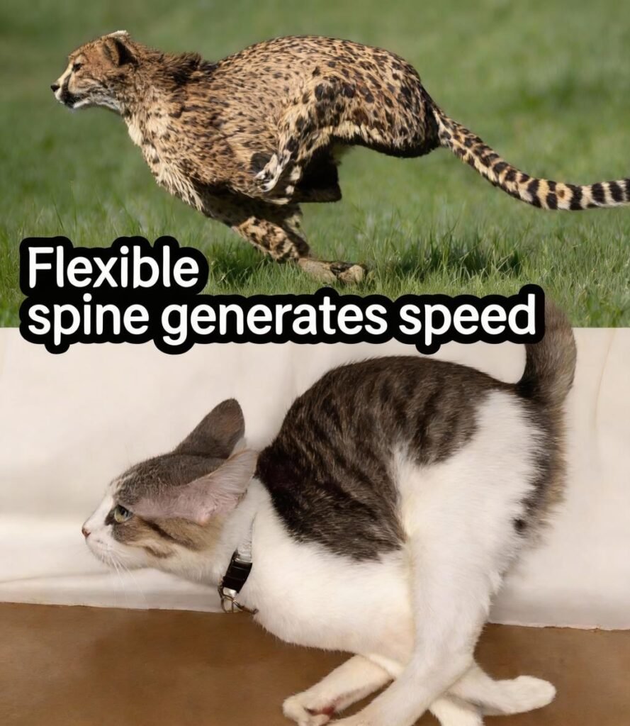
Cats’ flexible skeletons give them exceptional agility, allowing them to move with a grace reminiscent of cheetahs. The familiar “arched back” posture in cats is a result of their fully carnivorous diet. Compared to other animals, their intestines are relatively short, so the spine doesn’t need to support the internal organs as heavily, resulting in a highly flexible structure. This flexibility not only produces the characteristic arch of the cat’s back but also allows for a streamlined, cheetah-like form during rapid movement.
Additionally, the ligaments connecting bones and the intervertebral discs that cushion each vertebra are extremely supple. This flexibility enables cats to twist their bodies and groom almost all areas of their bodies—except the central back and parts of the face. Cats are naturally fastidious, and their glossy, well-groomed appearance is a direct result of this remarkable flexibility.
Interestingly, sex hormones appear to inhibit growth hormones. Cats spayed or neutered at an early age, when hormone secretion is interrupted, may experience slightly more unrestricted growth in their limb bones. Though this difference is subtle, it can result in marginally longer bones in the legs.
Head and Neck
Overview of Cat Head and Neck Anatomy
The following section covers the bones surrounding the head and neck of a cat.
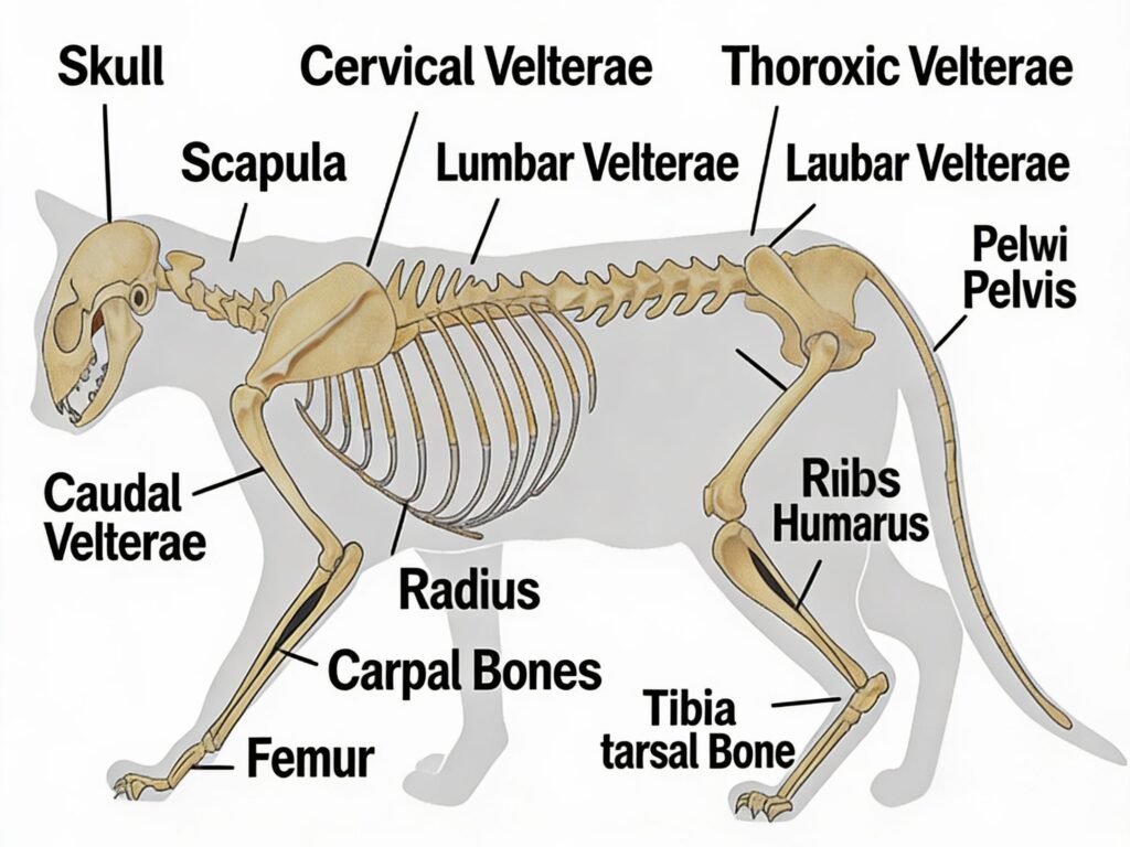
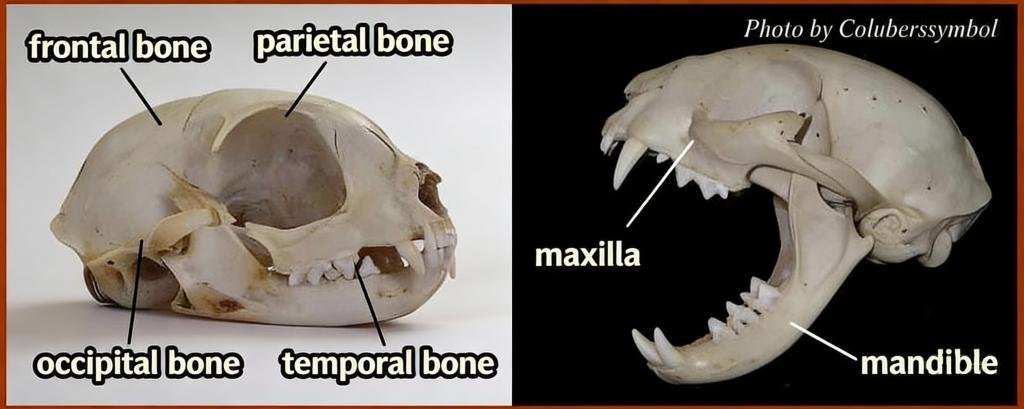
Skull
A cat’s skull consists of multiple bones and can be broadly divided into the cranial (braincase) portion and the facial portion.
The cranial portion is made up of eleven bones including the occipital, parietal, sphenoid, frontal, and temporal bones, which primarily protect the brain. When touching the head of a newborn kitten, you may notice it feels slightly soft. This is because the skull bones are not yet fully fused, allowing the kitten to pass more easily through the birth canal—a feature shared with human infants.
If intracranial pressure rises for any reason, causing the skull to deform, the condition is referred to as hydrocephalus.
The facial portion consists mainly of the mandible and maxilla, supporting the eyes, nose, and other sensory structures. Breeds like Exotic Shorthair, Persian, and Himalayan cats were selectively bred to maintain immature facial bones, resulting in the flat, “pushed-in” faces we recognize today.
Illustration: Cat skull showing parietal, occipital, frontal, temporal, mandible, and maxilla bones.
Cats’ faces are not elongated like some dog breeds (e.g., German Shepherds); they have a classic short-face structure. Despite their smaller skull volume:
- Their olfactory epithelium is smaller than that of dogs,
- They have 12 fewer teeth than dogs (adult dogs 42, adult cats 30),
…both of which are sufficient for their needs.
Brain Weight and Encephalization Quotient
A cat’s brain weighs approximately 27–32 grams, about 1% of its body weight.
The Encephalization Quotient (EQ) is calculated by dividing brain weight by two-thirds of body weight, multiplied by a scaling factor. EQ is often used as an indicator of intelligence, with higher scores generally associated with greater cognitive abilities.
Typical EQ values for various animals:
| Animal | EQ |
|---|---|
| Human | 7.4 |
| Dolphin | 4.8 |
| Chimpanzee | 2.2 |
| Crow | 1.25 |
| Dog | 1.2 |
| Cat | 1.0 |
Cervical Vertebrae
Cats have seven cervical vertebrae, just like humans, supporting the neck.
- The first cervical vertebra is called the atlas,
- The second cervical vertebra is called the axis,
…and together they allow the head to rotate smoothly. The atlas is the widest vertebra in the entire spine.
Torso / Trunk
The torso of a cat, spanning the chest to the lower back, consists of the thoracic vertebrae, ribs, and lumbar vertebrae.
Thoracic Vertebrae and Ribs
Cats have 13 thoracic vertebrae—one more than humans—and correspondingly 13 pairs of ribs.
The top nine pairs of ribs connect to the sternum, while the lower four do not.
The rib cage, supported by the intercostal muscles between each rib, facilitates breathing and protects the internal organs.
When you feel a cat’s chest, the bony ridges you detect are the sternum. Unlike humans, whose sternum is relatively flat, cats’ sternum is elongated, producing a more pronounced tactile sensation.
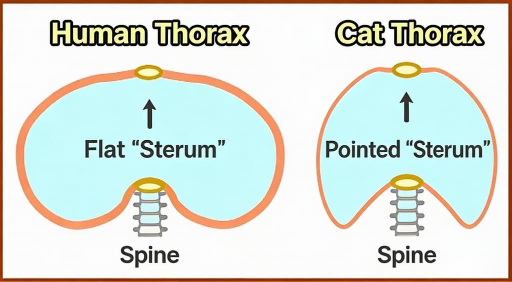
Viewed from above, a human thoracic cross-section is an oval extending sideways, forming a “chest plate” in the front. In cats, the oval extends front-to-back, creating what could be called a “chest ridge.”
Additionally, the 10th thoracic vertebra’s spinous process points backward, while the 11th points forward. This indicates the region between these vertebrae acts as a pivotal point for thoracic movement.
Lumbar Vertebrae
Cats have seven lumbar vertebrae—two more than humans. These extra vertebrae make the spine highly flexible.
This flexibility allows cats to twist their bodies mid-air when falling from heights, helping them land gracefully.
Unlike humans, whose lumbar spine is relatively straight for upright bipedal walking, a cat’s lumbar spine curves upward in a convex shape—a key feature of their body structure.
Between the lumbar vertebrae are intervertebral discs, which cushion impacts. Damage to these discs can lead to herniation, though it is less common in cats than in dogs.
Pelvis and Tail
Pelvis
The pelvis is made up of the ilium, ischium, pubis, and sacrum, forming a roughly hemispherical structure.
The sacrum consists of 3–4 fused bones at the base of the lumbar spine.
Like humans, the pelvis features a cup-shaped socket called the acetabulum, into which the femur fits snugly.
The cat’s hip joint, a synovial joint formed by the femur and acetabulum, is more prone to dislocation than other joints. However, strong, flexible ligaments hold the joint securely, allowing cats to move vigorously without injury.
Tail Vertebrae
Tail vertebrae, or caudal vertebrae, support the cat’s tail and generally number 18–19.
However, this varies widely: short-tailed breeds like the Manx may have only 4, while long-tailed cats may have up to 24.
Because of this variation, the total number of bones in a cat’s body is not fixed.
The Japanese Bobtail, a short-tailed breed from Japan, likely originated from natural mutations for shorter tails that were selectively bred over generations.
Tail vertebrae taper toward the tip, and the vertebral canal, which houses the spinal cord, usually ends around the ninth caudal vertebra. Beyond that, the canal becomes a shallow groove.
Forelimbs
Cats’ forelimbs, from shoulder to paw, are composed of the scapula, humerus, radius, ulna, and carpal bones.
Scapula
The scapula slides along the back ribs and connects forelimb muscles to the torso.
Like large cats, a walking cat’s scapula sits slightly above the spine.
Clavicle
The clavicle is small, located near the base of the neck, and serves as a muscle attachment point.
Unlike in humans, a cat’s clavicle floats, not connecting to other bones. This allows cats to squeeze their bodies through spaces as narrow as their heads.
Key muscles attached to the clavicle include the clavotrapezius, clavobrachialis, and cleidomastoid.
Humerus
The humerus is the main upper-arm bone. In humans, it supports the biceps. In cats, it mainly supports locomotion.
While the hindlimb’s femur is somewhat suspended, the forelimb humerus is always “engaged,” similar to holding a continuous push-up position.
Radius and Ulna (Forearm Bones)
The radius and ulna allow cats to rotate their paws to groom their faces.
This rotation is made possible by well-developed pronator and supinator muscles. Dogs lack this rotation and rely on their two forearm bones to absorb shock like springs.
Carpal Bones
The carpal bones form the wrist, consisting of several small bones connected by ligaments, dispersing impact forces.
Below the carpal bones are five metacarpals, ending in phalanges that contact the ground. Cats are digitigrade, walking on their toes.
Phalanges are divided into proximal, middle, and distal bones, with the distal phalanx bearing the claw. The distal phalanx acts like a pulley, enabling claw extension and retraction.
Hindlimbs
Cats’ hindlimbs, from thigh to paw, consist of the femur, tibia, fibula, and tarsal bones.
Femur
The femur is the thigh bone. Cats’ digitigrade gait keeps their knees bent while walking or running, giving the impression of walking in an “air chair” position.
The patella (kneecap) guides smooth leg movement. Displacement of the patella results in patellar luxation.
Tibia and Fibula
Together, these bones form the lower leg, acting like springs to absorb ground impact.
Tarsal and Metatarsal Bones
The tarsal bones, analogous to the human ankle, are multiple small bones connected by ligaments to distribute shock.
Below the tarsals are four metatarsals in the hindlimb, ending in phalanges that contact the ground.
Hind metatarsals are roughly twice the length of fore metacarpals, so cats maintain slightly bent hindlimbs to align with forelimb height.
This arrangement enables silent, stealthy movement, ideal for hunting.
When threatening another animal, cats fully extend their normally bent hindlimbs, raising their hips to appear larger.